Posterior Shoulder Tendon Anatomy : Shoulder Anatomy And Rotator Cuff Injury Body Complete. Together they assist in stabilizing the shoulder joint as well as in performing. Anatomy of shoulder hd wallpaper photographs anatomy of shoulder and neck pain on top of shoulder anatomy of shoulder bones The inner sleeve consists of two muscles of the rotator cuff, the infraspinatus, and the teres minor. (image courtesy of carolyn nowak, ann arbor, mich. The posterior segment attaches proximally along the inferior aspect of the scapular spine.
The main function of the triceps is the extension of the elbow joint. Together they assist in stabilizing the shoulder joint as well as in performing. All three segments attach distally to the deltoid tuberosity of the humerus via a common tendon. Axillary nerve (c5,6) from posterior cord of brachial plexus. You will also find coracoid process of scapula, subscapularis, teres major, serratus anterior, teres minor, supraspinatus, spine of scapula.
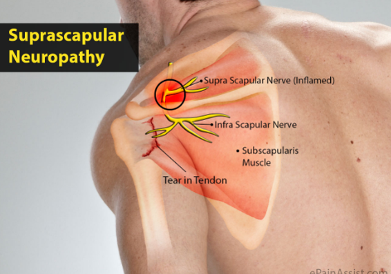
Exercises For Rotator Cuff Injury ð—£ ð—¥ð—²ð—µð—®ð—¯ from i0.wp.com Located superior to the shoulder joint, the deltoid muscle works with the supraspinatus to abduct the arm at the shoulder. The nerve itself is approximately 2.5 cm away from the glenoid rim and approximately 4 cm from the posterior corner of the spine of the scapula (plancher et al. The posterior aspect of the shoulder is covered by two muscular sleeves. Together they assist in stabilizing the shoulder joint as well as in performing. About eight shoulder muscles attach to the. A muscle contracts to move bones; The posterior deltoid is located on the back of your shoulder. This usually occurs secondary to repetitive use of the shoulder joint.
The shoulder joint, also known as the glenohumeral joint, is a ball and socket joint with the most extensive range of motion in the human body.
Rotator cuff tendonitis refers to inflammation of the tendons of the rotator cuff muscles. The posterior aspect of the shoulder is covered by two muscular sleeves. The main function of the triceps is the extension of the elbow joint. There are 10 muscles and 11 shoulder tendons related to shoulder mobility. You are in the emergency room when a patient is brought in, the loser in a street fight. In this image, you will find supraspinatus, deep muscles of the left shoulder anatomy, pectoralis major, deltoid in it. Limbs anatomy for android devices. The nerve itself is approximately 2.5 cm away from the glenoid rim and approximately 4 cm from the posterior corner of the spine of the scapula (plancher et al. The muscles of the shoulder have a wide range of functions, including abduction, adduction, flexion, extension, internal and external rotation. It also helps you raise and rotate your arm. Ebraheim's educational animated video describes muscle anatomy of the shoulder girdle and anatomy of the shoulder joint.anatomy of the shoulder muscles a. The nerve which passes through the quadrangular space of the posterior shoulder innervates which muscle? Its main function is shoulder extension, which is characterized by pulling your upper arms backward and bringing your shoulder blades together.
The rotator cuff is a collection of muscles and tendons that surround the shoulder, giving it support and allowing a wide range of motion. They also protect the main shoulder joint, the glenohumeral. (image courtesy of carolyn nowak, ann arbor, mich. Anterior fibers flex & medially rotate arm; Deltoid infraspinatus subscapularis supraspinatus teres major;
1 from Anatomy of shoulder components of the shoulder ideas posterior view of the shoulder high resolution wallpaper images: Together they assist in stabilizing the shoulder joint as well as in performing. The shoulder joint is composed of the glenoid (the shallow shoulder socket) and the head of the upper arm bone known as the humerus (the ball). The bursa is a small sac of fluid that cushions and. Anatomy of shoulder hd wallpaper photographs anatomy of shoulder and neck pain on top of shoulder anatomy of shoulder bones Shoulder muscle anatomy shoulder blade muscles body anatomy organs human body anatomy gross anatomy bicep tendonitis scapula muscular system anatomy anatomy images. Illustrations of (a) anterior and (b) posterior shoulder show supraspinatus (ss), infraspinatus (is), subscapularis (s), teres minor (tm), and long head of the biceps brachii tendon (b). The deltoid functions to flex, abduct, and extend a straight arm against resistance applied to the anterior, lateral, and posterior.
Your rotator cuff helps provide shoulder motion and stability.
Muscles, tendons, and ligaments combine to keep your arm bone in your shoulder socket. Your rotator cuff helps provide shoulder motion and stability. The main function of the triceps is the extension of the elbow joint. Posterior — the back of the shoulder medial — the side of the shoulder closest to mid body lateral — the side of the shoulder farthest from mid body proximal — located nearest to the point of attachment or reference, or center of the body The subscapularis, teres minor, supraspinatus, and infraspinatus. Together they assist in stabilizing the shoulder joint as well as in performing. The rotator cuff is a group of four muscles and tendons that surround the glenohumeral joint. (image courtesy of carolyn nowak, ann arbor, mich. The most commonly affected tendons in the shoulder are the four rotator cuff tendons and one of the biceps tendons. The posterior part of the deltoid muscle forms the outer sleeve of muscle. Anatomy, shoulder and upper limb, triceps muscle the triceps brachii is a large, thick muscle on the dorsal part of the upper arm. The rotator cuff is a collection of muscles and tendons that surround the shoulder, giving it support and allowing a wide range of motion. Located superior to the shoulder joint, the deltoid muscle works with the supraspinatus to abduct the arm at the shoulder.
The bursa is a small sac of fluid that cushions and. This muscle is targeted in movements like the dumbbell rear deltoid raise, face pull and bent over row. It often appears as the shape of a horseshoe on the posterior aspect of the arm. Your rotator cuff helps provide shoulder motion and stability. Anterior fibers flex & medially rotate arm;
Test Your X Ray Iq Deep Posterior Shoulder Pain Airrosti from www.airrosti.com The posterior segment attaches proximally along the inferior aspect of the scapular spine. Posterior — the back of the shoulder medial — the side of the shoulder closest to mid body lateral — the side of the shoulder farthest from mid body proximal — located nearest to the point of attachment or reference, or center of the body Relevant anatomy anatomic lesions associated with posterior shoulder instability involve injury to the posterior labrum, inferior glenohumeral ligament, and capsule. Posterior fibers extend & laterally rotate arm. Posterior view of the shoulder. Anatomy, shoulder and upper limb, triceps muscle the triceps brachii is a large, thick muscle on the dorsal part of the upper arm. The bursa is a small sac of fluid that cushions and. The biceps tendon begins at the top of the shoulder socket (the glenoid) and then passes across the front of the shoulder to connect to the biceps muscle.
The posterior part of the deltoid muscle forms the outer sleeve of muscle.
Illustrations of (a) anterior and (b) posterior shoulder show supraspinatus (ss), infraspinatus (is), subscapularis (s), teres minor (tm), and long head of the biceps brachii tendon (b). Posterior fibers extend & laterally rotate arm. The posterior segment attaches proximally along the inferior aspect of the scapular spine. The main function of the triceps is the extension of the elbow joint. Posterior view of the shoulder. The rotator cuff is a collection of muscles and tendons that surround the shoulder, giving it support and allowing a wide range of motion. The inner sleeve consists of two muscles of the rotator cuff, the infraspinatus, and the teres minor. Posterior labral detachment (the reverse bankart lesion) has been described in approximately 50% of patients. The nerve itself is approximately 2.5 cm away from the glenoid rim and approximately 4 cm from the posterior corner of the spine of the scapula (plancher et al. In this image, you will find supraspinatus, deep muscles of the left shoulder anatomy, pectoralis major, deltoid in it. Anatomy of shoulder hd wallpaper photographs anatomy of shoulder and neck pain on top of shoulder anatomy of shoulder bones Anatomy, shoulder and upper limb, triceps muscle the triceps brachii is a large, thick muscle on the dorsal part of the upper arm. The biceps tendon begins at the top of the shoulder socket (the glenoid) and then passes across the front of the shoulder to connect to the biceps muscle.
Posterior — the back of the shoulder medial — the side of the shoulder closest to mid body lateral — the side of the shoulder farthest from mid body proximal — located nearest to the point of attachment or reference, or center of the body shoulder tendon anatomy. 1 the central bony structure of the shoulder is the scapula, where all of the muscles interact.

0 Comments:
Posting Komentar